ul. Szewska 4/5
31-009 Kraków
tel. 012 421 2520
|
Rectoscopy includes the following range of examinations:
- diagnostic rectoscopy,
- rectoscopy with the collection of biopsy specimens,
- rectoscopy treatment.
What is rectoscopy?
The procedure.
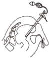
Rectoscopy is a test allowing for endoscopy of the anus canal and rectum with the use of rectoscope - rigid instrument about 20 - 30 cm long and 2cm in diameter (rectoscopes for children about 1cm in diameter). The optical fibers supply the light known as 'cold illumination' to the rectoscope.
What is the purpose of the examination?
Rectoscopy allows the specialists to assess the morphological condition of mucous membrane in the segment of large bowel. Moreover, the procedure allows doctors to collect the biopsy specimens for histopathological and bacteriological examination. The removal of polyps, foreign bodies and control bleeding is feasible to perform in this rectoscopy.
The indications as valid reasons to use the examination.
- Bleeding from the anus (also latent bleeding - the blood in the anus might be found only in the lab examination mainly because it cannot be observed with the naked eye).
- The pain in the anus and abdomen area.
- Some changes of the bowel emptying or the shape of stool (band-like stool, narrow stool), futile tenesmus or fecal incontinence.
- Tumour in the anus.
- The oozing of discharge from the anus.
- Pruritus of the anus.
- Other - the examination of the biopsy specimens.
Preceding procedures.
Examination per rectum
How to prepare for the examination?
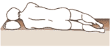
First of all, the doctor prepares the patient for the rectoscopy by carrying out the clysma - cleansing the anus with 1litre of warm water (temp. between 25° - 30° C). Enemas can be carried out when the patient lies down on the left side with the pad placed under the patient's buttocks (Picture 2-14). After the sterilization, the rectal nozzle for enema is connected with the irrigator by rubber tube and filled with water or solution recommended by the doctor. Afterwards, the clamp should be screwed on the nozzle which is later smeared with vaseline. The buttocks should be parted and then the rectal nozzle is inserted into the anus at a depth of 10 cm. After that, the irrigator should be raised - as high as the length of the tube allows the doctor to do it and then the clamp is released. When water flows down, the rectal nozzle is removed from the rectum. At that time, the patient turns on the right side and after few minutes there is bowel movement. The pad placed under the patient's buttocks should be taken out at the moment of tenesmus. The most appropriate time for rectocopy is 20-30 minutes after the final stage of bowel movement mainly because the faeces might flow later into rectum from the upper part of bowel. At present, there are ready-made clysmas (eg. Enema) available for faster bowel movement. It is not recommended to use oral laxatives. In some cases - eg. when there is a suspicion of ulcerating inflammation of large bowel - the rectoscopy might be performed without preparations because this allows the doctor to make an objective appraisal of mucous membrane lesions.
The examination is carried out with the local anaesthetization. It is also advisable to give the patient some tranquilizer.
Description of the examination.
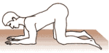
During the examination, the patient assumes a knee-elbow position and splays his/her legs. Those patients who are seriously ill or those with the lesions of their osseous and muscular system have the examination performed in the Sim's left-side position (patients keep their thighs close to each other, the buttocks are situated a little bit on the outside of the table with the turned trunk that the stomach is directed to the table).
At the beginning of the examination, the doctor observes the anus area and examines the rectum. After that, the ending of rectoscope lined with anaesthetic is inserted at a depth of 5 cm without the visual control of the rectum area. Finally, the obturator (gentle ending plug) is taken out from the rectoscope and allows to insert the instrument gently through the area of anal sphincter in order to carry on the observations.
The result of the examination is presented in the form of a description.
 |
 |
 |
|
The knee-elbow position during the rectoscopy.
|
The position of seriously ill patients who are not able to assume the knee-elbow position during the rectoscopy.
|
The image of rectoscopy.
|
Time
The examination usually lasts a few minutes.
Information that should be reported to specialist.
Before the examination:
- pain in the anus area,
- the effect of preparations for the examination (the effectiveness of the enema),
- occurrence of menorrhea.
During the examination: every painful complaints.
Complications after the examination.
On the whole, the examination is safe for the patient. Although, the perforation of the intestine hardly ever occurs, the complication might arise after the examination. Some patients might complain about small bleeding that usually subside by themselves.
|
|


